Biology and life cycle of Entamoeba histolytica
An overview of the life cycle, morphology, and development of Entamoeba histolytica.
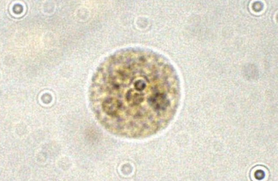
The infective cyst
The cyst is the non-motile, highly resistant infective form of Entamoeba histolytica. Measuring approximately 10 to 20 micrometers, a mature cyst houses four nuclei in a protective wall that permits survival in external moist environments for weeks. This wall also prevents digestion by stomach acid. Infection begins when mature cysts are ingested via contaminated food or water (2).
Image: Entamoeba histolytica cyst displaying 4 nuclei (5).
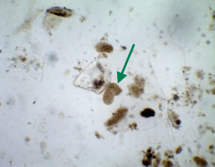
Excystation of active trophozoites
Upon entering the ileum, the cyst wall degrades, releasing trophozoites. Between 10 and 60 micrometers, these motile individuals colonize the cecum and large intestine. Trophozoites feed primarily on bacteria and cellular debris, adhering to the mucosal epithelial lining. Here, they may remain non-invasive or penetrate deeper into intestinal tissue and neighboring blood vessels (2). Blood smears from extra-intestinal cases may show ingested red blood cells inside the amoeba as well (9).
Image: Micrograph of Entamoeba histolytica trophozoite taken at 40x magnification (7). Note amorphous shape.
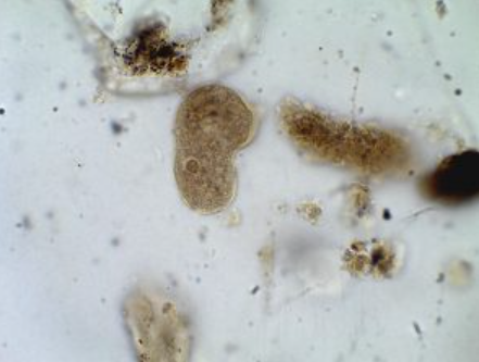
Encystation and transmission
Encystation protects the trophozoite as it feeds. Nuclear division proceeds twice within the developing cyst, forming four nuclei (2). The monogenetic life cycle halts when mature cysts are defecated, hardy against environmental stressors, until ingested by the next primary host, primarily humans, but occasionally domestic animals like dogs and cats (2, 8).
Image: Micrograph of Entamoeba histolytica trophozoite taken at 100x magnification (6). Note "Ground-glass" cytoplasm and single nuclei.
Create Your Own Website With Webador