Pathology & symptoms of Entamoeba histolytica
Explore the progression of infection from asymptomatic carriage to extra-intestinal disease.

Non-invasive colonization: asymptomatic carriage
In some cases, E. histolytica resides within the intestinal lumen without causing tissue damage. Asymptomatic carriers harbor an active protozoal colony and shed infective cysts in feces (2). They play a critical role in transmission while exhibiting no clinical signs of infection.
Invasive intestinal amoebiasis: acute colitis
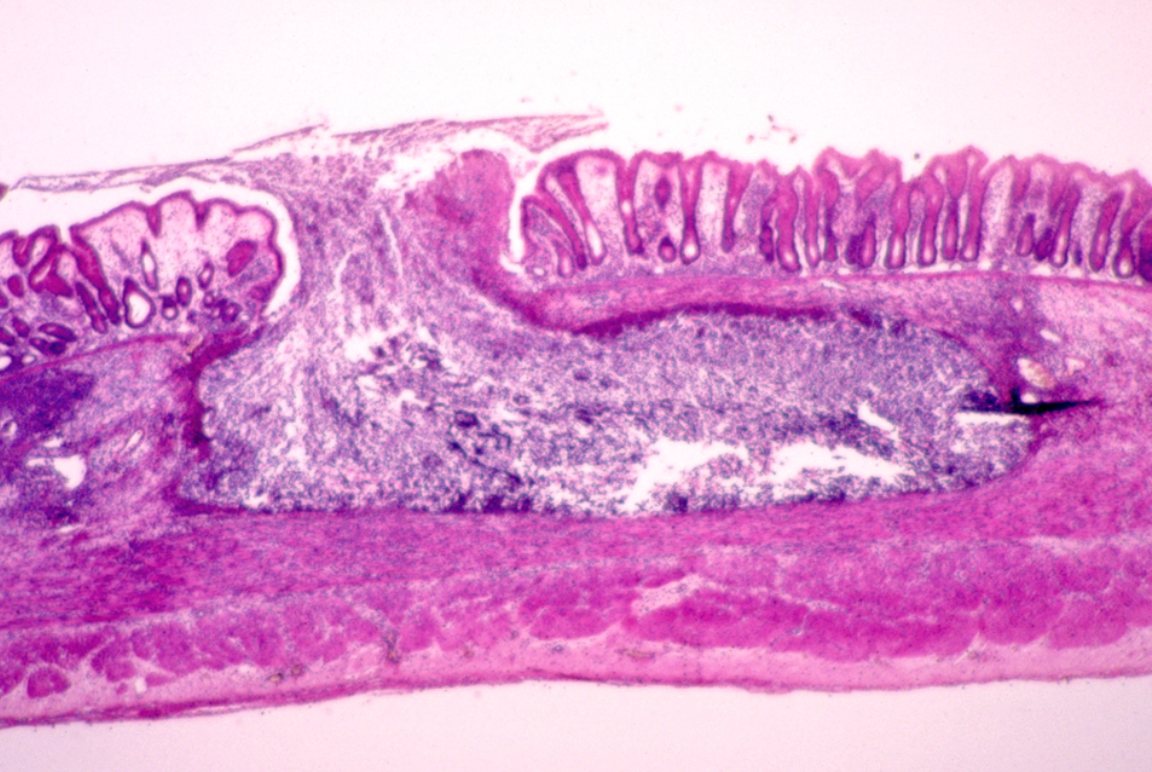
When trophozoites penetrate the mucosal lining, they cause flask-shaped ulcerations in the colon and cecum. Clinical presentation includes painful colitis, weight loss, and diarrhea that may or may not contain blood. Anemia may result from this (2, 8).
Image: "Flask-Shaped" ulcer in colon. Note death of the tissue within the ulcer, and the characteristic spreading of necrosis underneath the epithelium (9).
Extra-intestinal amoebiasis: metastasis
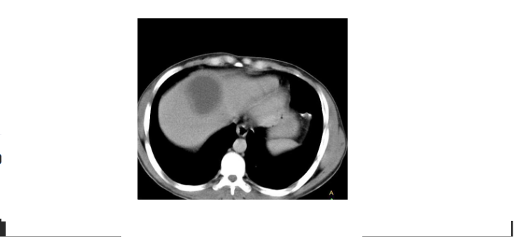
In severe cases, the parasite invades the bloodstream through the intestinal wall, most often forming liver abscesses or spreading to the lungs and brain. Patients present with right upper quadrant (RUQ) abdominal pain, sustained fever, apparent leukocytosis, and dramatic weight loss. The migration of the trophozoites from the digestive tract means this stage is not accompanied by blood loss and diarrhea (2).
Image: Computed tomography cross section depicting a large, round shadow in the liver caused by amoebiasis (4).
Create Your Own Website With Webador